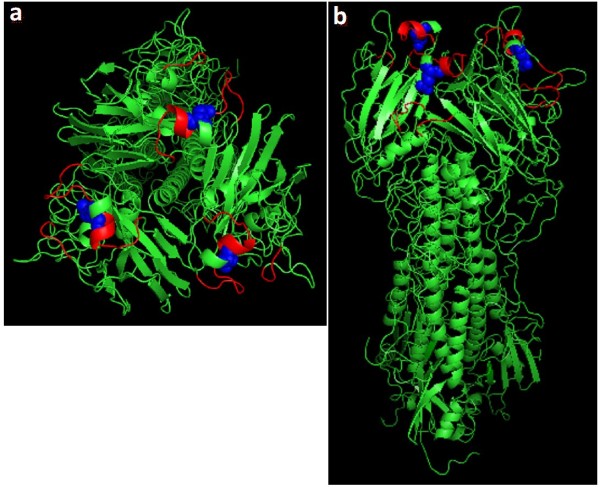
Figure 1.
HA structure with Asp to Ala mutation at residue 190 of the RBD. The 3D structure of the HA molecule was downloaded from Protein Data Bank webpage (http://www.pdb.org; 1HGG-A/Aichi/2/68 (H3)) and modified using the PYMOL Molecular Graphics System (DeLano Scientific, San Carlos, CA). a: top view of the HA molecule; b: side view of the HA molecule. Red: RBD. Blue balls: Residue 190 of the RBD.

