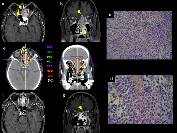
Figure 1.

Patient 2, a 70-year-old Japanese male with BCAC in the ethmoid sinus. (a) Axial contrast-enhanced T1-weighted MR image before C-ion RT, (b) Coronal contrast-enhanced T1-weighted MR image before C-ion RT, (c) Histological findings of HE staining at low-magnification, (d) Histological findings of HE staining at high-magnification, (e) Dose-distribution of C-ion RT in axial and coronal CT images, (f) Axial contrast-enhanced T1-weighted MR image 1 year after C-ion RT, (g) Coronal contrast-enhanced T1-weighted MR image 1 year after C-ion RT.
