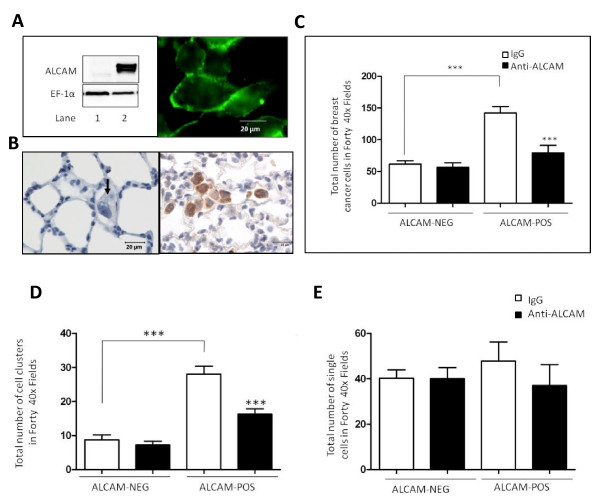
Figure 7.
ALCAM clusters tumor cells circulating through the lung. A) Ectopic expression of ALCAM in MDA-MB-435 tumor cells. Left panel; Western blot analysis of ALCAM in MDA-MB-435 clones transfected with an empty vector (lane 1) or, an ALCAM-GFP vector (lane 2). Protein loading was verified by probing the same western blot filter for the house keeping protein EF1-α. Right panel; Monolayer of MDA-MB-435-ALCAM-GFP cells were examined by live-cell microscopy to reveal expression of ALCAM at sites of cell-cell contact. B) Medium power image show virtual absence of control MDA-MB-435 cells, except for a single cell (arrow) while the ALCAM-expressing MDA-MB-435 clones form clusters (brown stain) after 90 minute perfusion in rat lungs. C) Number of tumor cells retained in rat lungs in experiments using ALCAM-positive (n = 7), or ALCAM-negative MDA-MB-435 cells (n = 4) pre-treated with non-immune IgG or monoclonal anti-ALCAM antibody. D) Number of tumor cell clusters retained in rat lungs in experiments using ALCAM-positive and ALCAM-negative MDA-MB-435 cells pre-treated with non-immune IgG (n = 7) or monoclonal anti-ALCAM antibody (n = 7). E) Number of single tumor cells retained in rat lungs in experiments using ALCAM-positive and ALCAM-negative MDA-MB-435 cells pre-treated with non-immune IgG (n = 7) or monoclonal anti-ALCAM antibody (n = 7).

