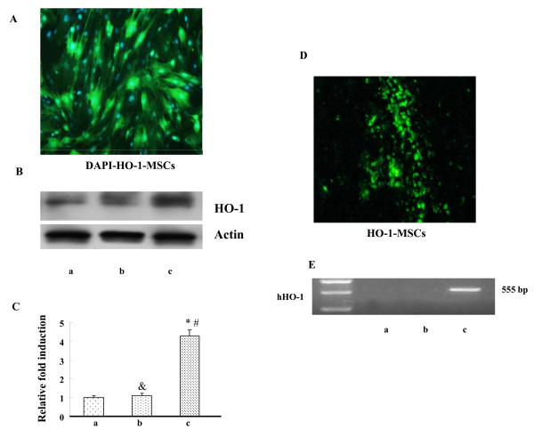
Figure 1.
HO-1 expression mediated by MSCs in Vitro and Vivo. (A) HO-1 expression mediated by MSCs with GFP in Vitro (200×). (B) Western blot analysis of HO-1 protein in MSCs with actin used as an internal control. Lane a, MSCs control (untransfected); lane b, Null-MSCs; lane c, Adv-HO-1-MSCs. (C) Graph showing the relative fold induction of HO-1 protein levels in MSCs, n = 6. * P < 0.05 compared with MSCs control (untransfected); &P > 0.05 compared with MSCs control (untransfected); # P < 0.05 compared with Null-MSCs. (D) Image from grafted HO-1-MSCs in the infarcted myocardium (200×). (E) RT-PCR detection mRNA in cardiac tissue. Lane a, MSCs control (untransfected); lane b, Null-MSCs; lane c, Adv-HO-1-MSCs.

