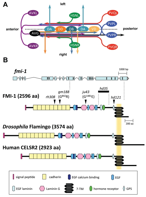
Fig. 1.
Development of the ventral nerve cord (VNC) in C. elegans, and gene model and protein domain organization of FMI-1. (A) The C. elegans VNC consists of two axon tracks with motoneuron cell bodies (light and dark orange, light and dark turquoise) lining the ventral midline (broken line). During embryogenesis, the AVG axon (black) pioneers the right VNC axon track. PVP axons (blue) decussate and extend on the contralateral side of the VNC. The PVPR axon pioneers the left VNC axon track. Both PVP axons are closely followed by PVQ axons (red). DA, DB and DD motoneurons (light and dark turquoise, dark orange, respectively) extend axons into the right axon track and send commissures circumferentially to the DNC. Later, AVK axons (purple) enter the VNC from the anterior. Postembryonically, VD motoneurons (light orange) differentiate and send processes into the VNC and towards the DNC. HSN axons (green) join the VNC at the vulva region. Command interneuron axons, which extend in the right axon track, are omitted for clarity. Ventral view. (B) The fmi-1 gene. The top part illustrates the exon-intron structure of fmi-1. The positions of alleles used in this study are mapped onto the protein structure (arrowheads, bold line). The protein domain organization of Drosophila Flamingo and human CELSR2 are shown for comparison.

