Fig. 4.
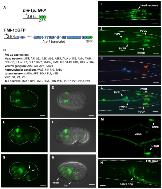
fmi-1 expression and localization. (A) Transcriptional and translational reporter constructs. (B) Cells expressing fmi-1p::GFP. Expression of the fmi-1p construct was detected embryonically (C-H) and postembryonically (I-N). (C,D) Gastrulation-stage embryo. (E,F) Comma-stage embryo. (G,H) 1.5-fold-stage embryo with expression in motoneurons (G, arrows). D, F and H are overlays of Nomarski and GFP channels. (I) L1 larva head region. (J-L) fmi-1p expression (green) in PVP (cyan) and PVQ (red) neurons. (M) Adult animal, vulva region. (N) FMI-1::GFP expression in the adult. Broken white lines indicate the outline of animals where possible. (C-H,N) Lateral views; (I-M) Ventral views. In all pictures, anterior is towards the left. Scale bars: 10 μm.

