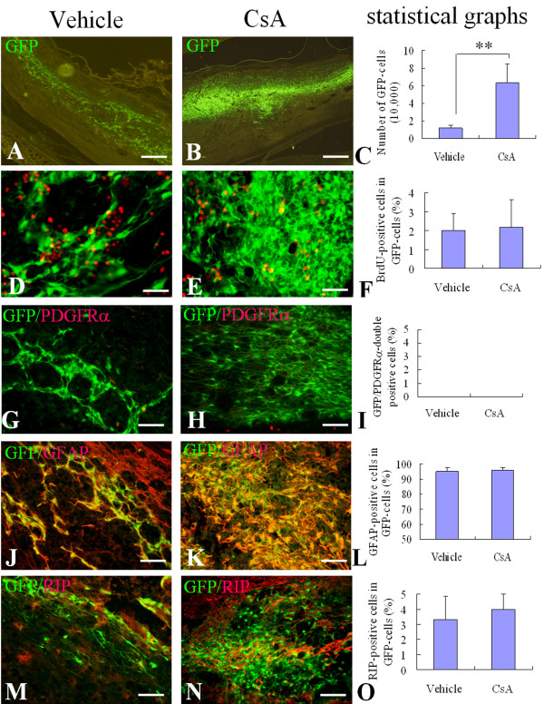
Figure 2.
Survival and differentiation of OPCs after engraftment into the injured adult rat spinal cord for 2 weeks. Two weeks after cell transplantation, the survival and differentiation of engrafted cells were detected in sagittal sections of injured spinal cord. A, B: Representative pictures show surviving GFP-cells in the vehicle (A) and CsA (B) treated groups. D, E, G, H, J, K: Representative merged images show GFP/BrdU-double positive cells (yellow) in the vehicle (D) and CsA (E) treated groups, GFP/GFAP-double positive cells (yellow) in the vehicle (G) and CsA (H) treated groups, and GFP/RIP-double positive cells (yellow) in the vehicle (J) and CsA (K) treated groups. The statistical graphs show the number of GFP-cells (C), the percentages of BrdU (F), GFAP (I) and RIP (L) positive cells in GFP-cells. Data are given as means ± SD, n = 6, ** p < 0.01 (t test). Scale bar in A, B: 500 μm; in D, E: 20 μm; in G, H, J, and K: 40 μm.

