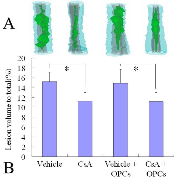
Figure 5.

Three-dimensional reconstruction of lesion volumes at the 7th week after contusive SCI. A: Representatives of the three-dimensional reconstruction of a 10 mm spinal cord segment from each group containing the lesion cavity (green). The spinal cord contours and white matters are shown in semitransparent blue, and the gray matter is depicted in gray. B: Data are given as means ± SD, n = 6, * p < 0.05 (ANOVA).
