FIG. 10.
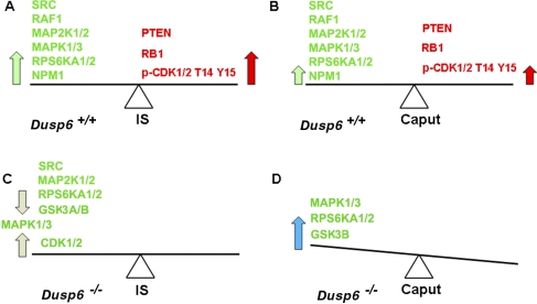
Diagram illustrating our working hypothesis of regulation of cell proliferation in the IS and caput before and after Dusp6 disruption. A) In the Dusp6+/+ IS, strong proproliferation activities of SRC, RAF1, MAP2K1/2, MAPK1/3, RPS6KA1/2, and NPM1 (large green arrow) balance strong antiproliferation activities of PTEN, RB1, and phosphorylation of CDK1/2 at T14 Y15 (large red arrow). B) In the Dusp6+/+ caput, the lesser proproliferation activities of SRC, RAF1, MAP2K1/2, MAPK1/3, RPS6KA1/2, and NPM1 (small green arrow) balance the lesser inhibitory force from PTEN, RB1, and phosphorylation of CDK1/2 at T14 Y15 (small red arrow). C) After loss of Dusp6 in the IS, the decreased proproliferation responses resulting from decreased SRC, MAP2K1/2, RPS6KA1/2, and GSK3A/B activities balance the increased proproliferation responses resulting from increased CDK1/2 activity (light gray arrows). MAPK1/3 activities were not affected by the loss of Dusp6. In the end result, cell proliferation regulation did not altered significantly in the Dusp6−/− IS, and the tissue size of Dusp6−/− IS was unchanged. D) Increased MAPK1/3, RPS6KA1/2, and GSK3B activities resulting from Dusp6 disruption (blue arrow) tipped the original balance to a proproliferation state in the Dusp6−/− caput, which resulted in a larger caput.

