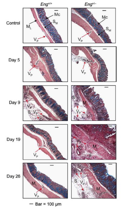
FIGURE 4.

Mucosal microvessels undergo more extensive angiogenesis in the distal colon of the DSS-treated Eng+/− mice. Mice were treated as described in FIGURE 1 and sacrificed as indicated. Distal colon sections were stained with Movat pentachrome to highlight the blood vessels. In the control groups, few vascular protrusions with one or more vessels (VP) from the submucosa (SM) through the muscle layer (ML) were observed. Vessels of increasing size and in larger numbers protruded from SM through ML and into the serosa (S), starting from day 5 and peaking at day 9 in the Eng+/+ mice; a recovery phase followed with VP showing a near normal pattern by days 19-26. In the Eng+/− mice, the vascular remodeling persisted beyond day 9 with a large number of VP lodging into the serosa at days 19-26. Bar = 100 μm.
