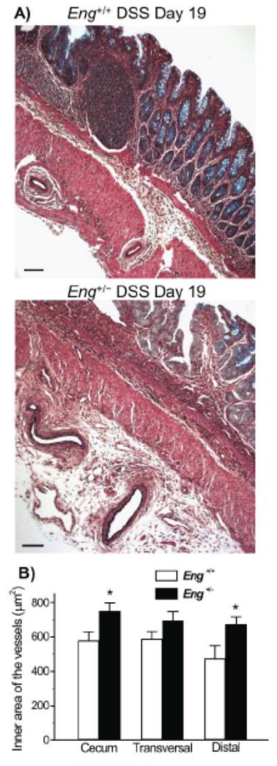
FIGURE 6.

Enlarged vessels are observed in the colon of DSS-treated Eng+/− mice during the chronic phase of colitis. A) Representative images of Movat pentachrome stained distal colon sections showing dilated vessels in the serosa of Eng+/− mice. In Eng+/+ mice, vessels were smaller and found in the muscle layer. Bar =100 μm. B) The inner area of all vessels (<60 μm) present in the sections of cecum, transversal colon and distal colon was measured. Vessels were significantly larger in cecum and distal colon respectively when comparing Eng+/− and Eng+/+ mice (*P < 0.05; N=5).
