Figure 1.
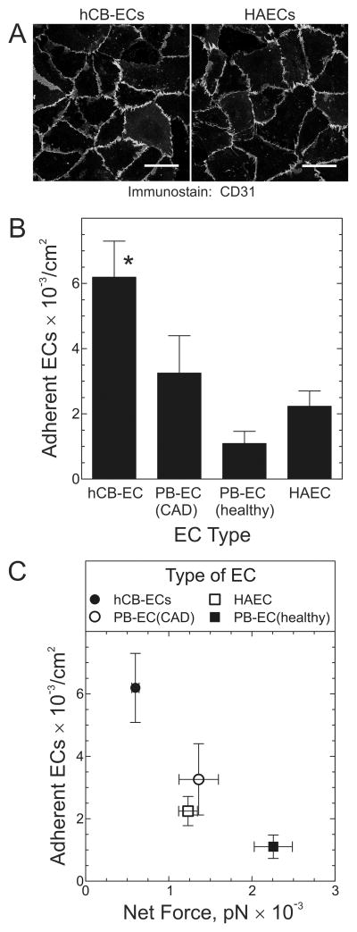
hCB-ECs demonstrate greater adhesion than HAECs or ECs derived from peripheral blood (PB-ECs). A, HAECs and hCB-ECs were immunostained for CD31 and imaged immunofluorescently. Shown are figures representative of ≥7 independent experiments. Scale bar = 50 μm. B, The indicated type of EC was suspended at 500,000 cells/ml and superfused for 10 min over quiescent SMCs to which was adsorbed exogenous fibronectin, collagen I and collagen III to augment adhesion (Supplemental Figure 1). The flow velocity used created shear stress of 0.5 dyn/cm2. The number of adherent ECs are plotted as the mean ± S.E. from 4 independent experiments. Compared with PB-EC(healthy) and HAEC: *, p < 0.05. C, The number of adherent ECs is plotted against the net fluid force acting on ECs exposed to 0.5 dyn/cm2. The net fluid force was calculated as described in Expanded Methods.

