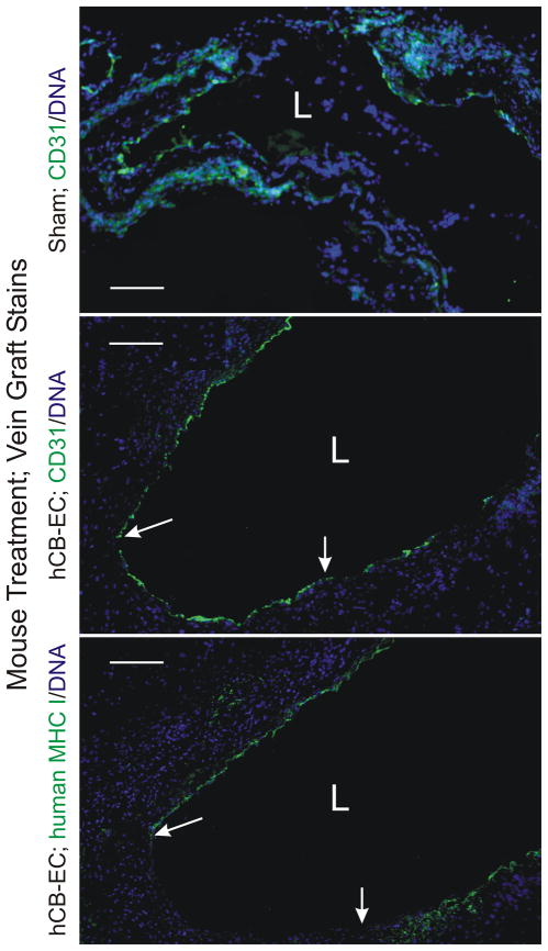
Figure 4.
Infusion of hCB-ECs enhances vein graft endothelialization. Vein grafts from mice with the indicated treatment were harvested 2 wk post-operatively, and serial cross sections were stained for DNA (Hoechst 33342) and either CD31 (PECAM), with IgG that recognizes both human and mouse CD31 (top two panels), or human (but not mouse) MHC I (bottom panel). The lumen is designated with an “L.” Between the arrows lies intimal surface covered by CD31-positive cells that are negative for human MHC I. Shown are individual vein grafts representative of specimens from eight hCB-EC-treated and 2 sham-treated mice (all other sham-treated mice had thrombosed grafts). Serial sections stained with isotype control IgG yielded no green color (not shown). Scale bars = 100 mm (original magnification × 110).

