
Figure 5. Effects of Asp3 domain disruption upon the export of GspB736flag.
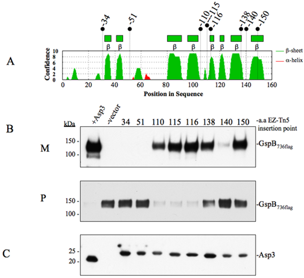
(A) Secondary structure analysis of Asp3. Locations of EZ-Tn5 insertions are shown above. (B) Western blot analysis of GspB736flag export from PS1244 derivative strains carrying in-frame insertions within Asp3. Culture media (M) and protoplasts (P) were collected from exponentially growing strains and prepared as described in the methods and materials. Proteins were separated by SDS-PAGE (3–8%) and following Western blot transfer, GspB736flag was probed with anti-flag antibody. (C) Western blot detection of Asp3 and the in-frame insertion mutants. Proteins from the protoplast fraction were separated by SDS-PAGE (4–12%) and probed with anti-Asp3 antibody.