Fig. 2.

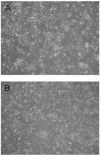
Phase-contrast photomicrographs of cultured primary hepatocytes. (A) Typical cubic hepatocytes shortly after isolation; (B) scattered, elongated hepatocytes after 48 hr (magnification, 40×).

Phase-contrast photomicrographs of cultured primary hepatocytes. (A) Typical cubic hepatocytes shortly after isolation; (B) scattered, elongated hepatocytes after 48 hr (magnification, 40×).