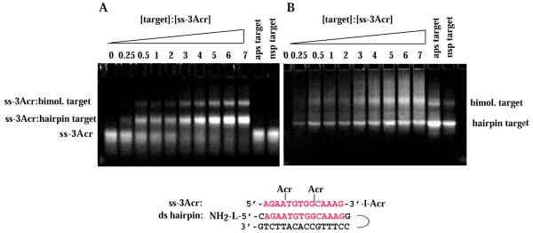
Figure 4.
Band shift assay of binding of ss-3Acr to the ds hairpin target. Conditions as in Figure 2. (A) Acridine fluorescence. (B) Fluorescence of multi-strand species after staining with EtBr. The concentration of ss-3Acr was 14 µM (strands) in all lanes. Concentrations of the ds targets are as in Figure 2.

