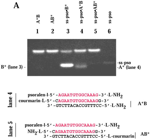
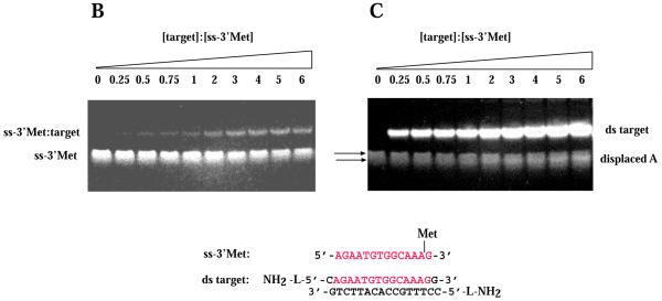
Figure 5.
Target strand displacement by ss-pso and ss-Met ODNs. (A) 5′,7-Diethylaminocoumarin-labeled A* or B* in the AB intermolecular target. ss-pso, 30 µM; ds AB, 140 µM. Lane 1, A*B duplex; lane 2, AB* duplex; lane 3, ss-pso·B* duplex with excess complementary B*; lane 4, ss-pso·A*B triplex; lane 5, ss-pso·AB* triplex; lane 6, ss-pso. (B and C) Titration of ss-3′Met with increasing concentrations of AB intermolecular target. (B) Methidium fluorescence. (C) Fluorescence of multi-strand species after staining with SybrGold. The concentration of ss-3′Met was 28 µM in all lanes. ds target concentrations are given as a factor of 28 µM (oligonucleotides) for each lane. Target lanes: lane 1, 0; lane 2, 0.25; lane 3, 0.5; lane 4, 0.75; lane 5, 1; lane 6, 2; lane 7, 3; lane 8, 4; lane 9, 5; lane 10, 6.


