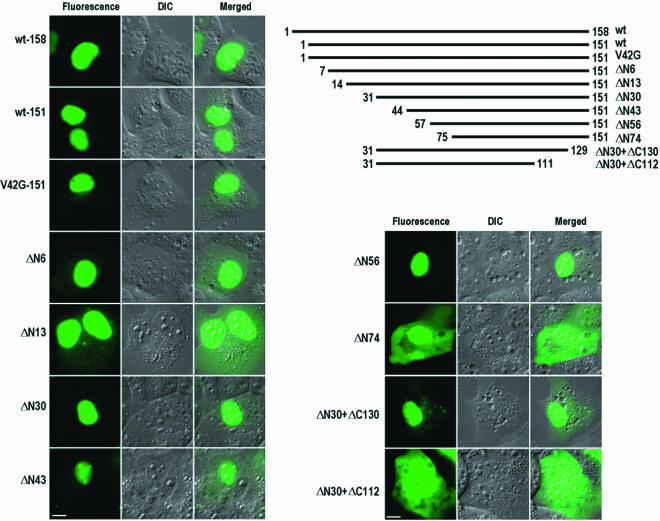
FIG. 5.
Mapping of NLSs in 16E6 in COS-1 cells. Three versions (158 aa, 151 aa, and 151 aa with V42G) of full-length 16E6 were compared for their subcellular localizations. The 151-aa version with V42G was similar to wild-type 151-aa 16E6 except for a V-to-G mutation at residue 42 due to an nt 226 5′ splice site mutation (GU to GG). 16E6 was truncated either from the N terminus to the C terminus or from the C terminus to the N terminus. The truncated E6-GFP fusions were expressed in COS-1 cells transfected with plasmids and imaged at 24 h after transfection. Numbers at the ends of the lines are positions of the first and last residues of the protein. DIC, differential interference contrast; wt, wild type. Scale bars, ∼8 μm.

