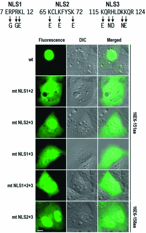
FIG. 7.
Mutational analysis of 16E6 NLS motifs in full-length 16E6 in COS-1 cells. Point mutations in the individual NLS motifs were the same as those described in the legends to Fig. 4 and 6. Cell images were captured at 24 h after transfection. DIC, differential interference contrast; wt, wild type; mt, mutant. Scale bar, ∼8 μm.

