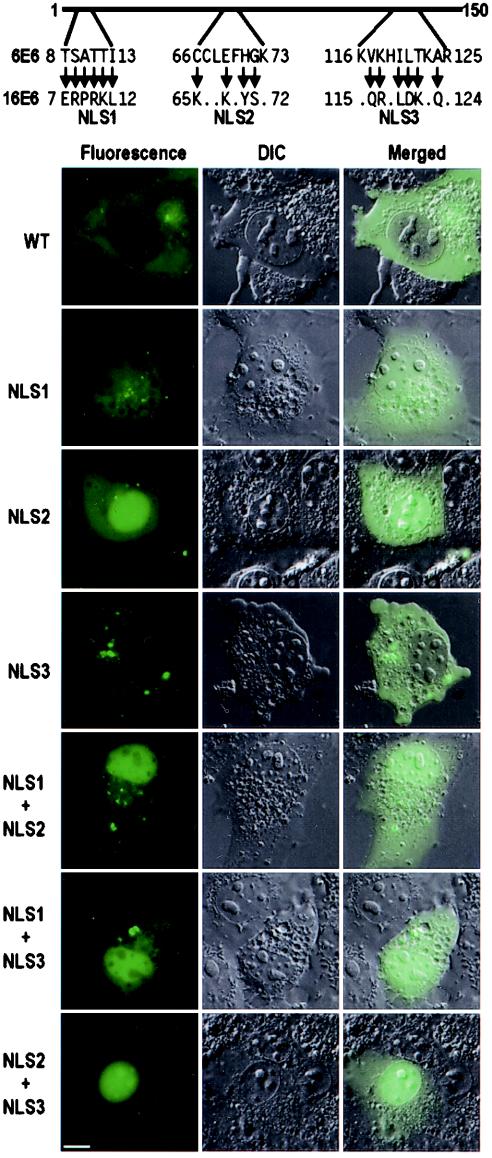
FIG. 8.
Conversion of a cytoplasmic 6E6 protein into a nuclear protein by 16E6 NLS motifs. The corresponding 16E6 NLS regions in 6E6 were replaced with individual 16E6 NLS motifs. The cellular distribution of the chimeric E6 protein with one or two 16E6 NLS motifs in COS-1 cells was imaged at 6 h after transfection. Arrows are as described in the legends to Fig. 4 and 6. DIC, differential interference contrast; WT, wild type. Scale bar, ∼8 μm.

