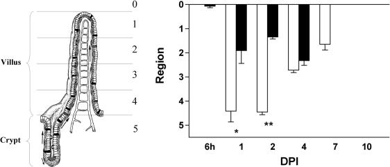
FIG. 4.
The pattern of epithelial vacuolization is more extensive than the pattern of replicating virus. Crypt-villus units in the jejunum were divided into five regions of equal length (see the left part of the figure). The position of the vacuolated cells (open bars) and virus-containing cells (solid bars) on the crypt-villus axis was scored from 1 to 5, representing the regions from the tips of the villi to the crypts. At 1 and 2 dpi, infected cells were exclusively found in the upper part of the villi, whereas vacuolated cells were observed along the entire villi and even at the base of the villi. Values are means + SEM (error bars). * and **, P < 0.01 and P < 0.001, respectively (Student's t test). At the left, a crypt-villus unit is shown and the different regions are indicated.

