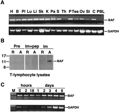
FIG. 1.
Expression of BAF mRNA in human tissues. (A) Agarose gel analysis of semiquantitative RT-PCR experiments done using two multiple tissue panels of first-strand cDNAs. Similar amounts of BAF cDNA (273 bp) were amplified from heart (H), brain (B), placenta (Pl), lung (Lu), liver (Li), skeletal muscle (Sk), kidney (K), pancreas (Pa), spleen (S), prostate (P), testis (Tes), ovary (Ov), small intestine (SI), and colon (C). BAF mRNA was not detected in thymus (Th) or peripheral blood leukocytes (PBL). Control experiments using primers specific to housekeeping enzyme GAPDH verified that all tissues amplified similar amounts of the 800-bp GAPDH fragment, confirming RNA integrity in these samples. (B) Western blot of protein lysates from resting (R) and day 4 in vitro-activated (A) CD4+ T lymphocytes probed with preimmune (Pre) or immune (Im) rabbit serum against human BAF. Monomeric BAF migrates at 11 kDa on gels (57). Recognition of BAF was specific, because no signal was obtained when immune antibodies were pretreated with peptide antigen (Im+pep). (C) Agarose gel analysis of semiquantitative RT-PCR experiments performed using RNA purified from CD4+ T lymphocytes at the indicated times after activation. M, molecular size markers.

