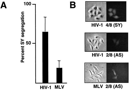
FIG. 3.
Frequency of SY segregation after infection early in G1. (A) Percentage of SY GFP colony patterns after infection of HeLa cell doublets, 3 h into G1 with HIV-1 or MLV GFP vectors. Standard deviations (error bars) are shown. (B) Representative patterns of GFP expression after infection of G1 cell doublets with the HIV-1 and MLV GFP vectors and subsequent synchronous outgrowth. Examples of AS and SY patterns are shown.

