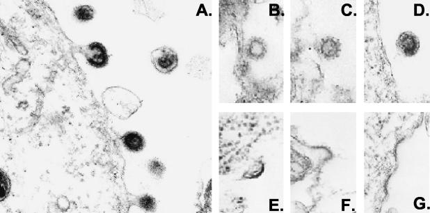
FIG. 2.
Electron microscopic analysis of viral budding. 293T cells were transfected with ACH in the absence or presence of 100 U of IFN/ml for 3 days. Cells were fixed, and viral particles were visualized by electron microscopy. Virus particles are seen budding through the plasma membrane of ACH-expressing cells in the absence of IFN (A) or the presence of IFN treatment (B to D). No virus particles were detected when cells treated with 50 μM proteasome inhibitor lactacystin for 2.5 h prior to harvest. Slightly curved and electron-dense thickenings are seen in panels E to G. Magnification, ×50,000.

