
Figure 4.
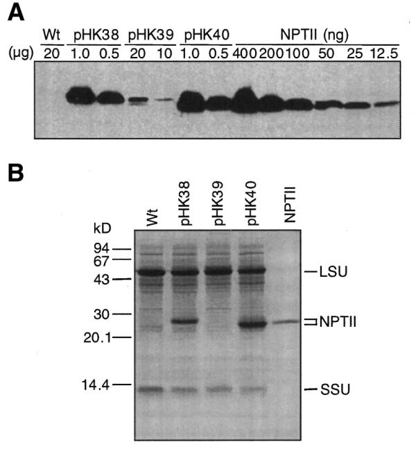
NPTII accumulation in tobacco leaves. (A) Immunoblot analysis to detect NPTII. The amount of TSP (µg) loaded on the SDS–PAGE gel is indicated above the lane. A NPTII dilution series was electrophoresed on the same gel. Lanes are marked with the plasmid name used for plant transformation. A protein sample from wild-type tobacco was also loaded (Wt). (B) Protein gel (20 µg/lane) stained with Coomassie Brilliant Blue R250. Control, NPTII (700 ng). The positions of NPTII and the Rubisco large (LSU) and small (SSU) subunits are marked.