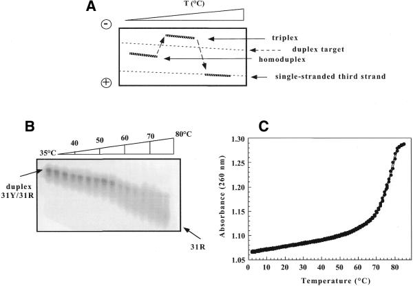
Figure 2.
(A) Model TGGE gel: a linear temperature gradient was applied perpendicular to the direction of the electrophoresis run. The arrows indicate the different species and their expected mobilities. (B) TGGE assay carried out with the 31R·31Y target duplex radiolabelled on the oligopurine strand at 2 µM in 50 mM HEPES (pH 7.2), 100 mM NaCl and 10 mM MgCl2. The linear temperature gradient covered the range 35–80°C. The arrows indicate the corresponding species. (C) UV melting and renaturation profile recorded at 260 nm of the 31R·31Y target duplex at 2 µM in a buffer containing 20 mM sodium cacodylate (pH 7.2), 100 mM NaCl and 10 mM MgCl2.

