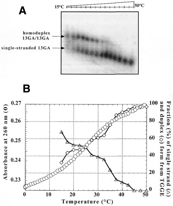
Figure 3.

(A) TGGE assay carried out with 20 nM radiolabelled 13GA oligonucleotide in the presence of 2 µM unlabelled 13GA in 50 mM HEPES (pH 7.2), 100 mM NaCl and 10 mM MgCl2. After overnight incubation at 10°C the sample was run on a TGGE gel with a linear temperature gradient between 15 and 50°C. The arrows indicate the corresponding species. (B) UV melting profile recorded at 260 nm of the homoduplex at 2 µM (circles) in a buffer containing 20 mM sodium cacodylate (pH 7.2), 100 mM NaCl and 10 mM MgCl2 was compared to the quantitative analysis of (A). Triangles, percentage of homoduplex as a function of the temperature; diamonds, percentage of single-stranded 13GA.
