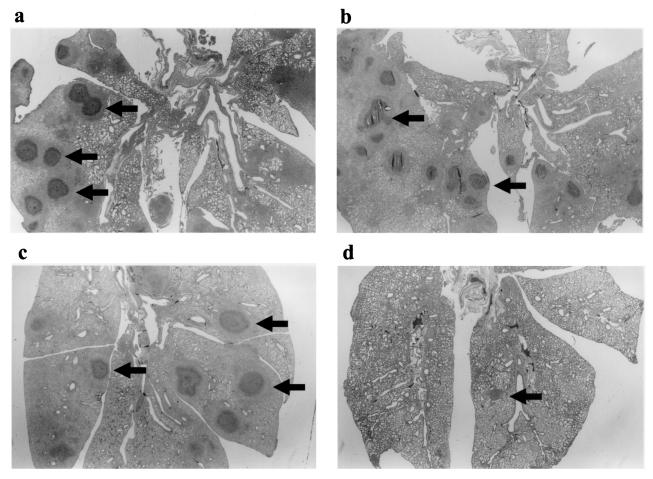
FIG. 2.
Histopathological examination of lung specimens from mice killed 10 days after treatment. Each specimen exhibited typical features of lung abscesses consisting of a central zone comprising a bacterial colony with infiltration of acute inflammatory cells (hematoxylin and eosin stain). Arrows show the lung abscesses. (a) control; (b) TEIC-treated group; (c) VCM-treated group; (d) DQ-113-treated group. Note that the severity of the inflammatory process is less in DQ-113-treated mice than in the mice in the other groups. Magnifications, ×5.

