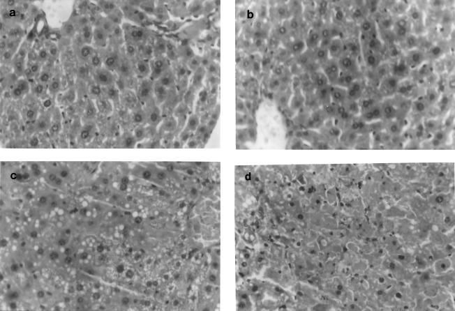
FIG. 2.
Liver histopathology (methylene blue and eosin yellow staining) indicates normal biliary parenchyma in control (a) and triclosan-administered (b) mice. Liver sections of infected mice (c) show hydropic and fatty changes with areas of hemorrhage, indicative of severe liver damage, which is considerably reduced in infected mice administered triclosan (d).

