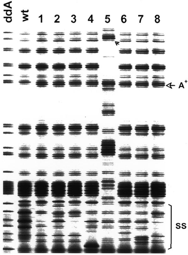
Figure 4.

Screening of the signature sequence to demonstrate independence of identical mutations. The partial fluoroimage shows both the variation of terminating band patterns in the signature sequence element (SS) and eight identical mutations that occurred at bp 172 as indicated by arrows. Although most banding changes in the signature sequence element were caused by nucleotide substitutions, sample 5 also involved a multi-nucleotide insertion that led to concomitant upward banding shifts. Terminating fragments shown in this image were generated from one of the DNA strands by primer PS189JR700 in a limiting dATP reaction. The results from the complementary strand are not shown.
