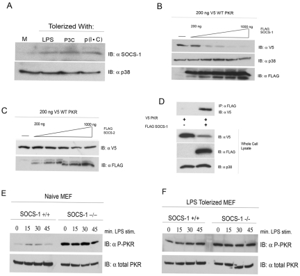
FIG 5 .
SOCS-1 physically interacts with and negatively regulates PKR. (A) Primary peritoneal macrophages were stimulated for 18 h with medium (M), LPS (10 ng/ml), P3C (100 ng/ml), or p(I ⋅ C) (10 µg/ml). Cells were harvested, and the levels of SOCS-1 protein were examined by Western analysis. (B and C) HEK293T cells were transfected with 200 ng V5-tagged WT PKR and either empty vector or an increasing amount of cDNA expressing FLAG-tagged SOCS-1 (B) or SOCS-2 (C). Twenty-four hours following transfection, whole-cell lysates were subjected to Western analysis with antibodies against the indicated species. (D) HEK293T cells transfected with V5-PKR alone or in conjunction with FLAG-tagged SOCS-1. Cell lysates were immunoprecipitated with anti-FLAG antibody and separated by SDS-PAGE, followed by Western blotting with the indicated antibodies. These data are representative of 3 independent experiments. (E and F) WT and SOCS-1−/− MEFs were cultured overnight in medium alone (E) or in medium supplemented with LPS (100 ng/ml) (F). Following 18 h of treatment, cells were washed and restimulated with LPS (250 ng/ml) for the indicated times. Whole-cell lysates were resolved by SDS-PAGE and probed with antibodies against phosphorylated or total PKR. These data are representative of 3 independent experiments.

