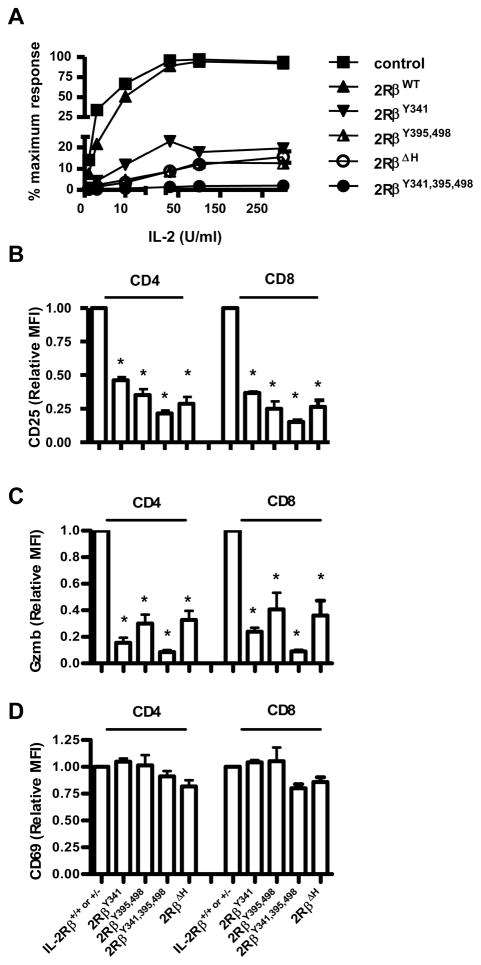
Figure 5. Proliferative and functional responses by T blasts bearing mutant IL-2Rβ.
Spleen cells from the indicated mice were culture with anti-CD3 for 48 hr. (A) The activated T cells were washed and re-cultured in the indicated level of IL-2 for 24 hr. 3H-thymidine was added during the last 4 hr of culture. Each data point represents the mean of at least 3 separate experiments, where the highest response was normalized to 100%. The activated CD4+ and CD8+ spleen cells were stained for (B) CD25, (C) granzyme B (Gzmb), or (D) CD69. The MFI of staining by control IL-2Rβ+/+ or +/− was normalized to a value of 1 and used to compare staining by T cells bearing mutant IL-2Rβ. Data are the mean ± SEM of ≥3 mice/group <16 wks of age.

