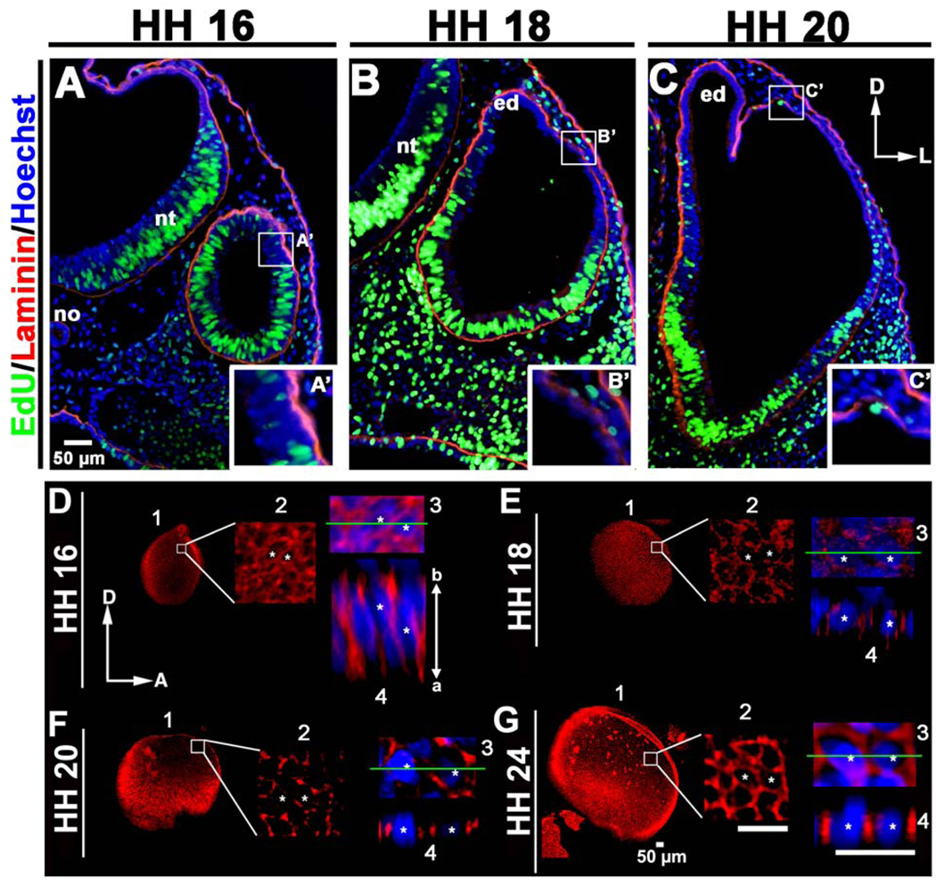
Figure 2. Increased cell proliferation does not drive expansion of the chick dorsolateral otocyst.
(A–C) Transverse sections of embryos pulsed for 2 hours with EdU (green), counterstained with anti-laminin (red) and Hoechst stain (blue) showing that the region that will undergo dorsolateral expansion (boxed, HH 16), or was undergoing dorsolateral expansion at the time the embryo was collected (boxed, HH 18 and 20). The dorsolateral otocyst contains few EdU-positive (i.e., mitotic cells in the S-phase of the cell cycle) cells, as compared to other regions of the developing otocyst, indicating that cell proliferation is not increased in the dorsolateral otocyst during its rapid expansion. D, dorsal; L, lateral; no, notochord; nt, neural tube; ed, endolymphatic duct. (A’-C’) Enlargements of the boxes in A–C showing epithelial thinning in the dorsolateral otocyst during its expansion. (D–G) Confocal imaging showing that E-cadherin (red; cell nuclei labeled with Hoechst staining, blue) is broadly distributed along the entire apicobasal (a–b) axis of the dorsolateral epithelium at HH 16, and then fragments as dorsolateral epithelial cells undergo thinning during HH 18–24. 1, panel showing the entire otocyst. 2, boxed area in panel 1 enlarged. 3, area in panel 2 containing two cells marked with asterisks enlarged. 4, panel showing a lateral view of the epithelium at the level of the green line in panel 3. D, dorsal; A, anterior.

