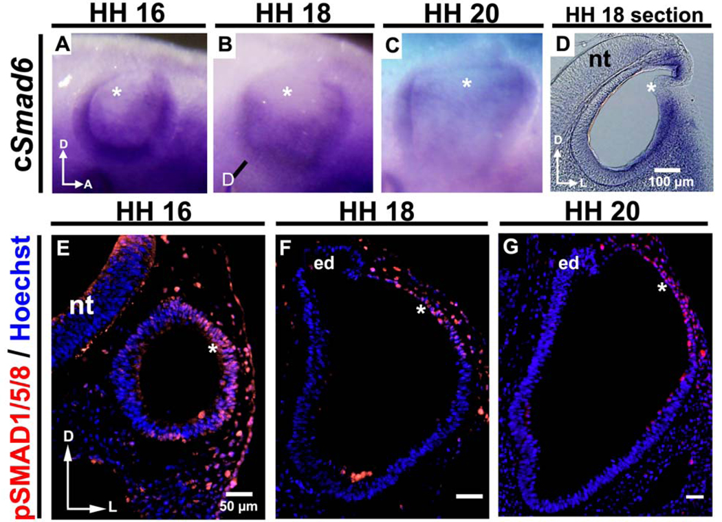
Figure 3. Smad6, an inhibitor of BMP signaling, is expressed only in the chick ventrolateral otocyst during its thinning and expansion, whereas pSMAD1/5/8 is expressed in the dorsolateral otocyst, indicating that this portion of the otocyst receives and responds to BMP signaling.
(A–C) As shown by whole mount in situ hybridization, Smad6, an inhibitor of BMP signaling, is expressed in the otocyst (but not in its dorsolateral wall; asterisks) during HH 16–20 when rapid thinning and expansion occurs (HH 16: n = 6; HH 18: n = 18; HH 20: n = 16). The line in (B) marks the level of the section shown in (D). Sections (D) reveal more clearly that expression occurs only in the ventrolateral wall of the otocyst, not in its dorsolateral wall (asterisks). D, dorsal; A, anterior; L, lateral; nt, neural tube. (E–G) Transverse sections (labeled with pSMAD1/5/8 antibody [red] and Hoechst stain [blue]) showing that pSMAD1/5/8 is expressed in the dorsolateral otocyst (asterisks; HH 16: n = 9; HH 18: n = 10; HH 20: n = 7), in contrast to Smad6, during HH 16–20. D, dorsal; L, lateral; nt, neural tube; ed, endolymphatic duct.

