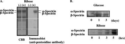
FIGURE 1.
Glycation of erythrocyte ghosts membrane and purified spectrin. Erythrocyte ghosts membranes were prepared by hypotonic lysis and incubated with ribose at 37 °C for up to 6 h. A, glycated ghosts membrane proteins were separated on 9% SDS-polyacrylamide gel and stained with CBB (left panel); a Western blot of a replicate gel probed with the anti-pentosidine antibody is shown (right panel). B, purified spectrin dimers prepared from unglycated erythrocyte membranes were incubated with glucose (upper panel) or ribose (lower panel) for the indicated periods of days or hours, respectively.

