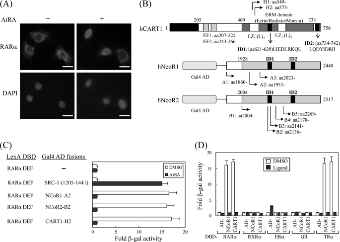
FIGURE 1.
Isolation of CART1. A, shown is subcellular localization of RARα in TM4 cells. Mouse RARα in TM4 cells were visualized by staining with rabbit anti-RARα polyclonal antibody and Texas Red-conjugated anti-rabbit antibody in the absence and presence of ligand AtRA. Each bar represents 10 μm. B, shown are schematic representations of human CART1, NCoR1, and NCoR2 (SMRTe) isolated by yeast two-hybrid screening. Locations of identified clones and functional domains are indicated: EF, EF-hand motif; LZ, leucine zipper; ID, nuclear receptor interaction domain. C, interaction of CART1 with hRAR(DEF) is shown. Interactions were monitored by introducing LexA DBD-fused RAR(DEF) and Gal4 AD-fused test partners in yeast strain L40 and by β-galactosidase (β-gal) assays with transformed yeast extracts. -Fold β-galactosidase activity indicates relative value compared with the Gal4 AD empty control. In all experiments, 1 μm AtRA was added to the yeast culture. DMSO was used as a control for AtRA. Data are shown as the averages of three independent experiments (mean ± S.D.). D, shown is interaction of CART1 with other nuclear receptor family members. Yeast two-hybrid assays were performed with the other NRs indicated in the presence of their cognate ligands: RAR, 1 μm AtRA; RXR, 1 μm 9-cis RA; ER, 1 μm estradiol (E2); GR, 5 μm deoxycorticosterone; TR, 1 μm triiodo-l-thyronine (T3). Data are the average of three independent experiments (mean ± S.D.).

