
Table 3.
Chemical Structures and Images of 17 Positives Identified in Dengue Virus Infection Assay
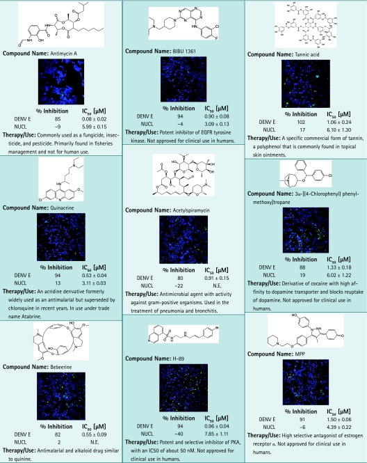 |
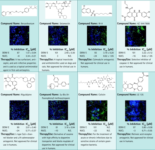 |
Images of cells tested with compounds at 2.5 μM are shown. Primary screening data as % inhibition and subsequent dose–response results are shown. For each data point, average and standard deviation calculated from duplicate data points are given. Plates were fixed and stained as described in the Materials and Methods section. Images were acquired using INCA3000, with green channel for DENV E and blue channel for detection of Hoechst-stained nuclei.
EGFR, epidermal growth factor receptor; N.E., no effect.