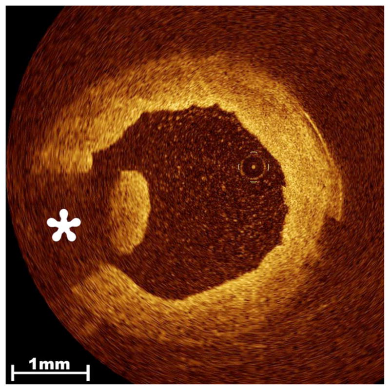
Fig. 5.

Thrombus in the endoscopically harvested SV. Residual clot strands that remain within the lumen of the procured vessel are readily detected by OCT imaging and range in severity from a single minute strand to near occlusive thrombus. Clot appears as an intraluminal lobulated mass with high signal intensity that produces characteristic radial signal attenuation (asterisk) due to the presence of entrapped red blood cells.
