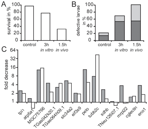
Figure 4. Comparison of in vitro and in vivo egg aging.
(A–C) Eggs were fertilized immediately, with a 3-hour delay (in vitro) or from a batch obtained 1.5 hours later (in vivo) from the same female shown also in Fig. 3 as exp. 8. The survival rate (A) and the percentage of defective development (B) observed at the free swimming larval stage 40 [21] is given. The defective development includes malformation (dark grey) as well as undersized and retarded development (light grey). (C) From each experiment the aging induced decrease of the fourteen polyadenylated transcripts analyzed in Fig. 2 is given (gray boxes: in vitro aging; white boxes in vivo aging).

