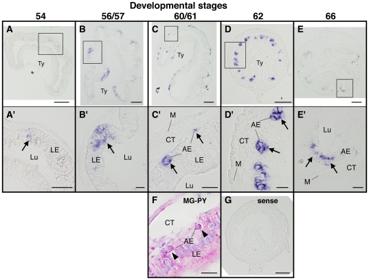
Figure 5. Spatiotemporal expression of LGR5 mRNA in the small intestine during natural metamorphosis.
Cross sections of the intestine at premetamorphic stage 54 (A, A′), prometamorphic stage 56/57 (B, B′), metamorphic climax stages 61 (C, C′, F) and 62 (D, D′, G), and the end of metamorphosis (stage 66) (E, E′) were hybridized with LGR5 antisense (A–E′) or sense probe (G). To compare the localization of LGR5 mRNA (C, C′) with that of adult epithelial progenitor cells, the serial sections at stage 60/61 were stained with methyl green-pyronin Y (MG-PY) (F). Arrows indicate the cells expressing LGR5 (A′–E′), while arrowheads indicate adult epithelial progenitor cells (F). Higher magnification of boxed areas in (A)–(E) are shown in (A′)–(E′). Sense probe did not produce any signal (G). Note that at metamorphic climax stage 61, LGR5 mRNA was localized in the islets between the larval epithelial cells and the connective tissue (C, C′). These islet cells were identified as the adult epithelial progenitor cells strongly stained red with pyronin Y (F) [29]. AE: adult epithelial cell including progenitor/stem cell, CT: connective tissue, LE: larval epithelial cell, Lu: lumen, M: muscle layer, Ty: typhlosole. Scale bars are 100 µm (A–E, G) and 20 µm (A′–E′, F), respectively.

