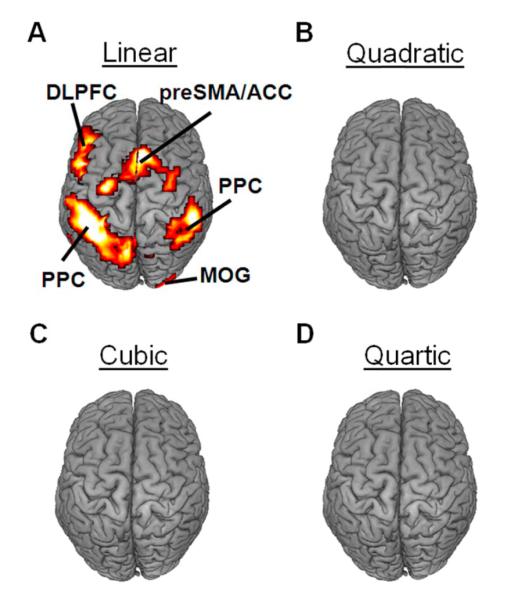
Figure 2.
Stimulus-evoked BOLD activity varied with RT in a predominantly linear fashion. (A) A linear relationship between stimulus-evoked activity and RT accounted for a significant proportion of signal variance in the bilateral posterior parietal cortex (PPC), the left dorsolateral prefrontal cortex (DLPFC), the pre-supplementary motor area / anterior cingulate cortex (preSMA/ACC), and the middle occipital gyrus (MOG). In contrast, quadratic (B), cubic (C), and quartic (D) relationships between stimulus-evoked activity and RT did not account for significant proportions of signal variance in any brain regions. All activations are overlaid on a 3D rendering of the MNI-normalized anatomical brain.

