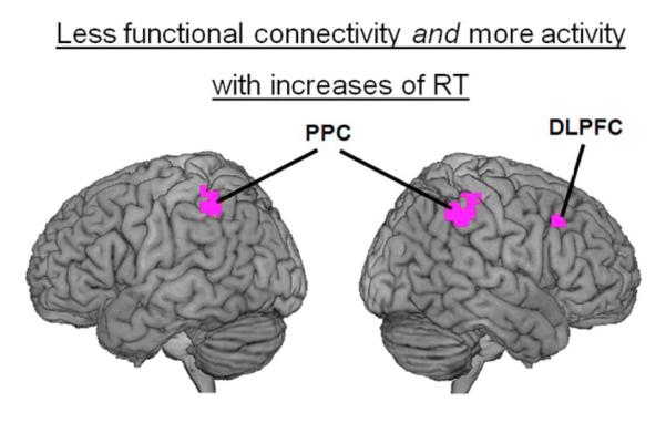
Figure 5.
Overlapping regions of the attentional network were implicated in the activity and functional connectivity analyses. Increases of RT were associated not only with increases of activity in the right dorsolateral prefrontal cortex (DLPFC) and in bilateral regions of the posterior parietal cortex (PPC), but also with reductions of functional connectivity between each of these regions and the ACC. These regions are overlaid on a 3D rendering of the MNI-normalized anatomical brain.

