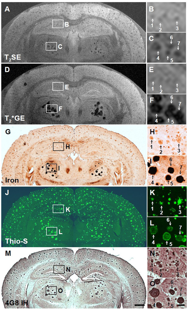
Fig. 3.
Five-way anatomic spatial co-registration of a 24-month old APP/PS1 AD transgenic mouse brain. Ex vivo MRI scans of matched sections imaged using either a (A) T2SE or (D) T2*GE pulse sequence. Matched adjacent histological sections processed with (G) DAB-enhanced iron staining, (J) thioflavine S amyloid staining, or (M) anti-Aβ peptide immunohistochemistry. Scale bar = 500 µm. (B, E, H, K, and N) Higher magnification of hippocampal plaques positively matched by spatial co-registration. Corresponding plaques are labeled with numbers when present in a particular section. (C, F, I, L, and O) Higher magnification of thalamic plaques positively matched by spatial co-registration. (O) Scale bar = 100 µm.

