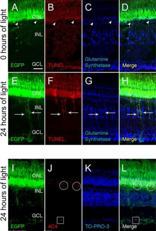
Figure 2. Müller glia engulf dying photoreceptor cell bodies in the ONL.
The Tg(XlRho:EGFP)fl1 transgenic fish express EGFP in rod photoreceptors from the Xenopus rhodopsin promoter. At 0 hours of light (A–D), control retinas exhibited EGFP expression (A and D, green) only in rods in the ONL (arrowheads) and the outer segments. There was no detectable TUNEL labeling (B) and low levels of Glutamine synthetase was restricted to the Müller glia (C and D). After 24 hours of light, EGFP was detected in both the ONL and INL (E and H, arrows), as was TUNEL labeling (F and H, arrows). Glutamine synthetase, which is expressed in Müller glia (G and H), colocalized with the rod-derived EGFP and TUNEL signal in the INL (H, arrows). An adjacent tissue section to that of panels E–H revealed that mAb 4C4-positive microglial/macrophage cells (J and L) were detected in both the ONL (circles) and GCL (boxes). These microglial/macrophage cells were EGFP-positive (I and L) in the ONL, but not in the GCL. The nuclear layers were identified by TO-PRO-3 staining (K and L). GCL ganglion cell layer; INL, inner nuclear layer; ONL, outer nuclear layer. Scale bar represents 25 μm.

