
Fig. 1.
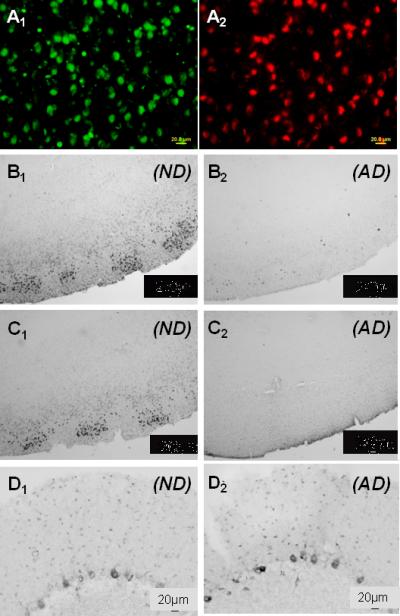
Representative micrographs of 5-methylcytosine and 5-methylcytidine immunoreactivity in entorhinal cortex layer II and cerebellar neurons from AD and ND cases. A1) High power micrograph of entorhinal cortex labeled with an antibody to 5-methylcytosine. A2) Same field labeled with an antibody to neuron-specific enolase to show co-localization (ND case). B1) Low power micrograph of 5-methylcytosine immunoreactivity in entorhinal cortex of an ND case to show general staining pattern. B2) 5-methylcytosine, AD case, entorhinal cortex. C1) 5-methylcytidine, ND case, entorhinal cortex. C2) 5-methylcytidine, AD case, entorhinal cortex. D1) 5-methylcytosine, ND case, cerebellum. D2) 5-methylcytosine, AD case, cerebellum. Staining with 5-methylcytidine in AD and ND cerebellum was virtually indistinguishable from that with 5-methylcytosine (see supplementary Fig. 1).