Figure 1.

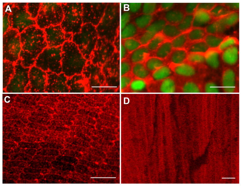
Confocal micrographs of αA-crystallin expression in the mouse lens. (A, B) Lens epithelial whole mounts prepared from adult wild-type mouse lenses were fixed and then incubated with a monoclonal antibody specific to αA-crystallin, followed by incubation with an Alexa568-conjugated secondary antibody (red); nuclei were stained with TOTO-1 (green). Cells were imaged by confocal microscopy at both apical (A) and basal (B) domains. Cells in the central epithelium are shown. (A) Note the expression of αA-crystallin on regions of plasma membrane at apical cell-cell interfaces. (B) Nuclei (green) are localized to the basal aspects of the lens epithelium where αA-crystallin was primarily cytoplasmic. Adult mouse lenses, ~150-day-old, were used to prepare lens epithelial whole mounts. (C) Cross-sectional and transverse sections of cortical lens fiber cells in adult wild-type mouse lenses. Cross-sectional slices were cut in the lens equatorial plane. Sections were stained with a monoclonal antibody to αA-crystallin and an Alexa568-conjugated secondary antibody (red). Intense staining for αA-crystallin was observed at the fiber cell interfaces in the outer cortical, hexagonally packed fiber cells. The periphery of the lens is at the bottom of the image. (D) In transverse sections the staining at cell-cell interfaces was shown to extend all along the lateral cell-cell borders of the cortical fiber cells. Cytoplasmic staining also was seen, which increased in the central fiber cells. Distance from the center of the lens is 400 μm. Scale bars = 10 μm (A), 20 μm (B, C), and 5μm (D).
