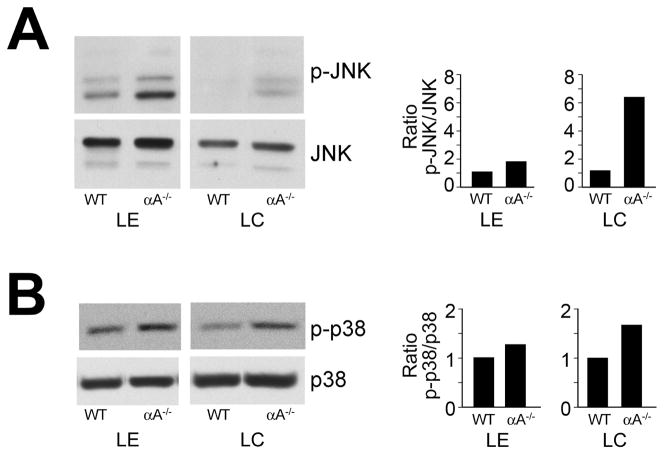
Figure 5.
Activation of JNK and p38 stress pathways in αA−/− lens epithelial and fiber cells. Lens epithelial (LE) and cortical fiber cell (LC) fractions were dissected from 18–20 wild-type (WT) or αA−/− mouse lenses. Cell extracts were examined by immunoblot analysis with antibodies to (A) p-JNK and JNK, and (B) p-p38 and p38. Note that while p38 and JNK were activated in lens epithelial and cortical fiber cells of αA−/− mice, the degree of activation of these stress kinases was much greater in the cortical fiber zone of αA−/− lenses. Protein bands were quantified, and ratios of activated to total protein were determined and presented as fold-change between αA−/− and WT lenses. The results are representative of three independent experiments.

