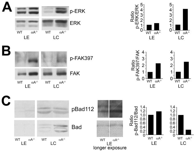
Figure 6.
Changes in survival signaling in αA−/− lens epithelial and fiber cells. Lens epithelial (LE) and cortical fiber cell (LC) fractions were dissected from 18–20 wild-type or αA−/− mouse lenses. Cell extracts were examined by immunoblot analysis with antibodies to (A) p-ERK and ERK1/2, (B) p-FAK397 and FAK, and (C) pBad112 and Bad. The results show increased activation of both ERK and FAK in αA−/− lens epithelial and cortical fiber cells, but decreased phosphorylation of Bad in the cortical fiber zone. Protein bands were quantified, and the ratios of activated to total protein were determined and represented as fold-change between αA−/− and WT lenses. The results are representative of three independent experiments.

