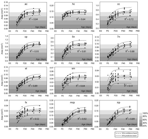
Figure 5.
Changes in white matter cross-sectional areas in twelve major white matter tracts. The area data were fitted to a sigmoidal model and the 95% confidence intervals are shown in the figures, with figure background indicative of the percentile of the estimated adult values (parameter a in Table 3). Structural abbreviations are: ac: anterior commissure; hc: hippocampal commissure; cc: corpus callosum; cp: cerebral peduncle; 5n: the trigeminal nerve; 7n: the facial nerve; st: stria terminalis; sm: stria medularis; fr: fasciculus retroflexus; fx: fornix; mcp: middle cerebellar peduncle; icp: inferior cerebellar peduncle.

