
Figure 2.
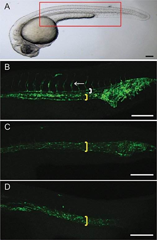
Dorsomorphin blocked intersegmental vessel (ISV) formation. (A) Normal embryo at 30 hpf (hours post fertilization) with red box illustrating the location where the confocal images were taken. Representative confocal z-stack images of Tg(fli1a:egfp)y1 zebrafish. (B) Normal with dorsal aorta (white bracket), posterior cardinal vein (yellow bracket) and ISV (white arrow) highlighted. Zebrafish exposed to 10 µM dorsomorphin (C) or 1 µM SU5416 (a pan vascular endothelial growth factor-a receptor inhibitor) (D) from 12 hpf have loss of the dorsal aorta and ISVs and expansion of the posterior cardinal vein. All are lateral views with anterior to left. Scale bars indicate 200 µm; n= 60–80 each group.